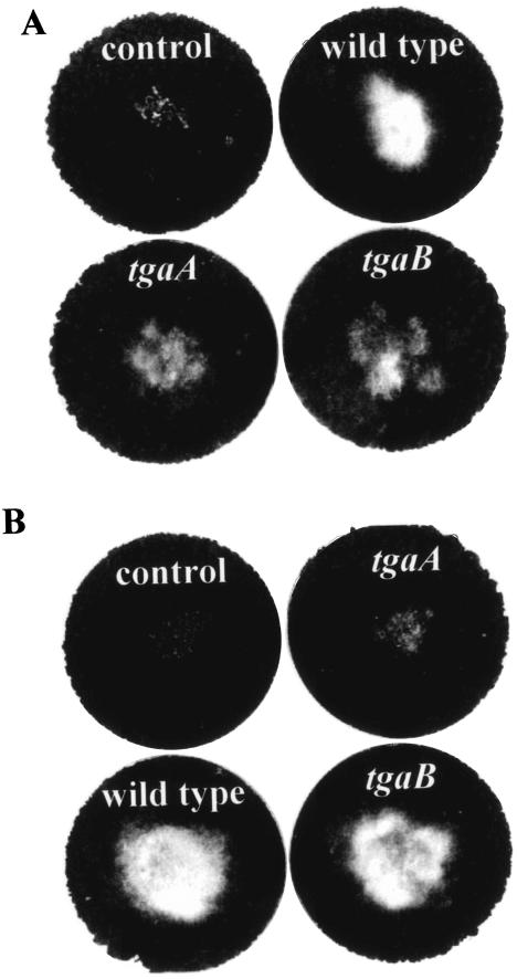
FIG. 6.
Parasitism of sclerotia by T. virens. The sclerotia were put in the middle of a soil plate after mixing of conidia into the soil. Control plates, sclerotia only, with no Trichoderma conidia. Plates were photographed after 7 days of incubation. Mycelial growth is visible as a light-colored colony contrasting with the dark background of the soil. (A) Growth on R. solani sclerotia. (B) Growth on S. rolfsii sclerotia.

