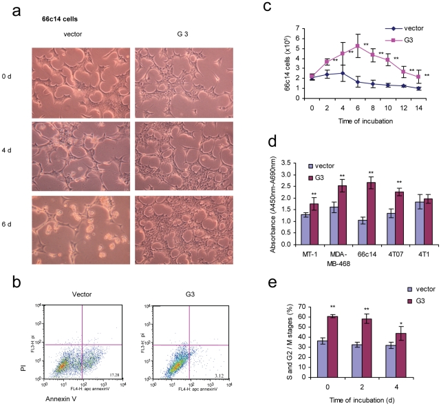
Figure 1. Versican G3 domain enhanced tumor cell survival in serum free medium.
a) G3-transfected and vector-transfected 66c14 (2×10⁵) were cultured in 10% FBS/DMEM medium in culture dishes for 12 hours. After cell attachment, we changed the medium to serum free DMEM and cultured cells for 6 days. Cell viability was analyzed by light microscopy. b) After culturing in serum free medium for 2 days, cells were analyzed with Annexin V and propidium iodide staining using flow cytometry. Annexin V and propidium iodide assays confirmed that cell death was apoptosis. c) G3- and vector-transfected 66c14 (2×10⁵) were cultured in 10% FBS/DMEM medium in culture dishes for 12 hours. After cell attachment, we changed the medium into serum free DMEM medium and cultured them for 14 days. Cells were harvested and counted under light microscopy every 2 days. Experimental results are compared with vector control group, n = 6, * p<0.05, **p<0.01, analyzed with t-test. d) 1×10⁴ G3- and vector-transfected human breast cancer cells MT-1 and MDA-MB-468, mouse mammary tumor cells 66c14, 4T07, and 4T1 were inoculated and cultured in 10% FBS/DMEM medium in 96 well culture dishes for 12 hours. After cell attachment, we changed the medium to serum free DMEM and cultured them for 8 days. WST-1 Cell Survival Assays were used to test cell viability. Compared with vector control group, n = 6, * p<0.05, **p<0.01, analyzed with t-test. e) Cell cycles were analyzed by flow cytometer. Compared with vector control group, n = 4, * p<0.05, **p<0.01, analyzed with t-test.

