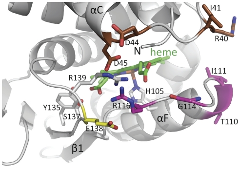
Figure 1. Close-up view of the heme-binding region of a homology model of the heme domain of sGC.
The heme (green) and its H105 ligand are depicted. Residues targeted for mutagenesis in the region between the αB-αC helices are colored brown, targeted residues in the αF-β1 strand region are shown in magenta and the flanking E138 residue in yellow. The αC helix, β1 strand, and the αF helix containing the heme-liganding residue H105 are labeled. Residues Y135, S137, and R139 as part of the YxSxR heme binding motif are shown in grey with the residues labeled.

