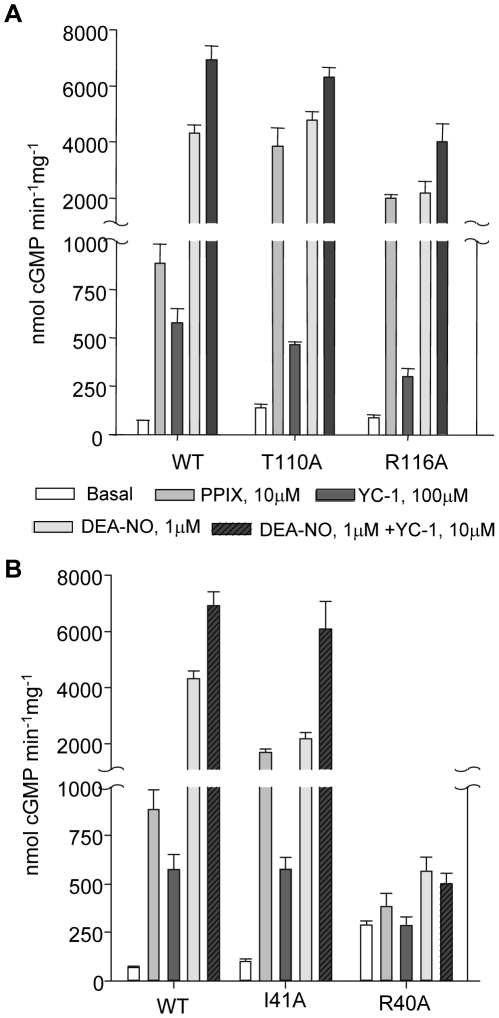
Figure 3. Basal and stimulated activities of purified WT and mutants.
3A: In the αF-β1 region, basal activity of α1/β1T110A as well as PPIX activation of α1/β1T110A and α1/β1R116A were significantly higher than WT. 3B: In the αB-αC loop, mutant α1/β1I41A exhibits a significantly higher response to PPIX and significantly lower response to DEA-NO compared to WT, yet sensitivity to DEA-NO +YC-1 was comparable to WT. α1/β1R40A lost the ability to respond to any activators. Measurements of activities were done at least three times on 50 ng of two to three independently semi-purified WT and mutants with each assay done in duplicate under each condition. Results are expressed in nmol cGMP. mg−1.min−1 ± S.E.M. Values and fold stimulation are provided in Table S1.

