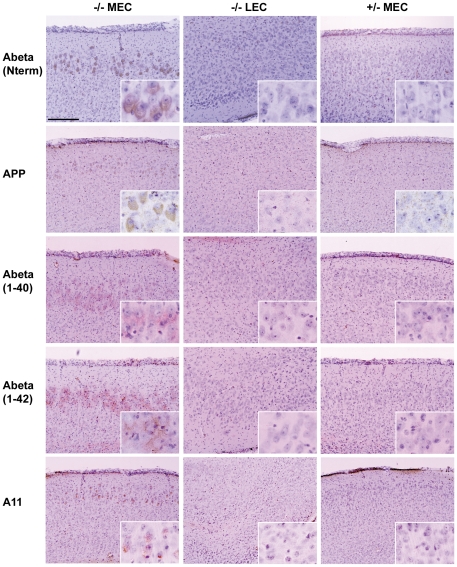
Figure 5. Elevated level of beta amyloid in the MEC region of MPS IIIB mouse brain.
The left column shows staining in the MEC region of MPS IIIB (−/−) mice, with the middle and right columns showing a control region (LEC) in the same mice and the MEC region in control (+/−) mice, respectively. The mice were 3 months old for Abeta (N term), 6 months old for APP, Abeta 1–40, Abeta 1–42, and 10 months old for A11. The antibodies used, from top to bottom, were: polyclonal antibody to amino acids 1–14 of amyloid beta, polyclonal antibody to amyloid precursor protein (APP), monoclonal antibody to peptide Abeta 1–40, monoclonal antibody to Abeta 1–42, and polyclonal antibody to A11.

