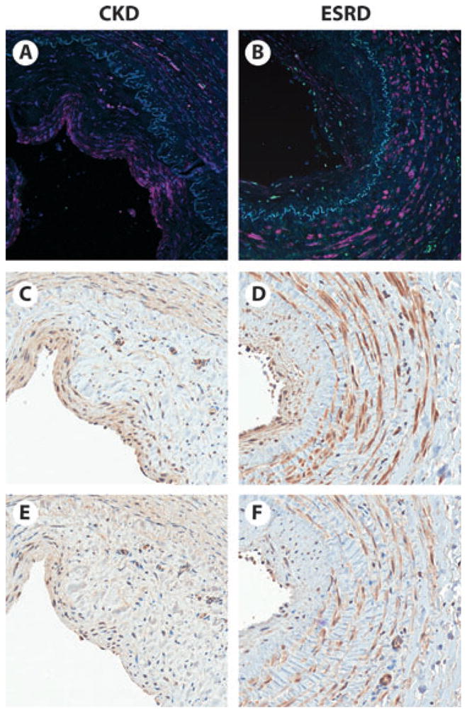
Fig. 2.

Serial vein segments from the cephalic vein of a pre-dialysis CKD (A, C, E) and an end-stage renal disease (ESRD) (B, D, F) patient. Immunoflourescence (A, B) shows abundant chymase (pink) expression in the thickened vessel intima and media (separated by elastic lamina, blue) of both the CKD and ESRD patient. Immunohistochemistry shows localization of TGF-b. (C, D) (brown) and IL-6 (E, F) (brown) primarily within the intima and media, in a similar distribution as chymase.
