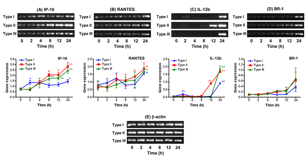
Figure. 1.
Effects of parasite strain type on the kinetics of pro-inflammatory cytokine and anti-apoptotic gene expression in WT microglial cells infected with T. gondii. Cells were infected with type I (GT1-FUDR3.3), type II (ME49 B7), or type III (CTG ARA-SYN) strains of T. gondii for 2, 4, 8, 12, and 24 h. RT-PCR amplification and analysis of gene expression of IP-10 (A), RANTES (B), IL-12b (C), Bfl-1 (D), and β-actin (E) were performed as described in section 2.3. Graphs represent levels of gene expression following normalization of the densitometric values of DNA bands to β-actin. Data points represent the means ± standard errors of the mean of three experiments. *, P < 0.05; **, P < 0.01 compared to uninfected cells (0 h time point).

