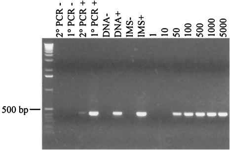
FIG.1.
The oocyst detection limit (oocysts per gram of feces) was determined by spiking fecal samples with decreasing numbers of oocysts. Secondary PCR products are shown after electrophoresis on a 1.2% agarose gel stained with ethidium bromide. From left to right, the lanes are as follows: molecular size standards; negative controls (−) for secondary (2°) and initial (1°) PCRs, respectively; positive (+) controls for 2° and 1° PCRs, respectively; negative and positive controls for DNA extraction, respectively; negative and positive controls for IMS, respectively; and fecal samples spiked with 1, 10, 50, 100, 500, 1,000, and 5,000 oocysts, respectively.

