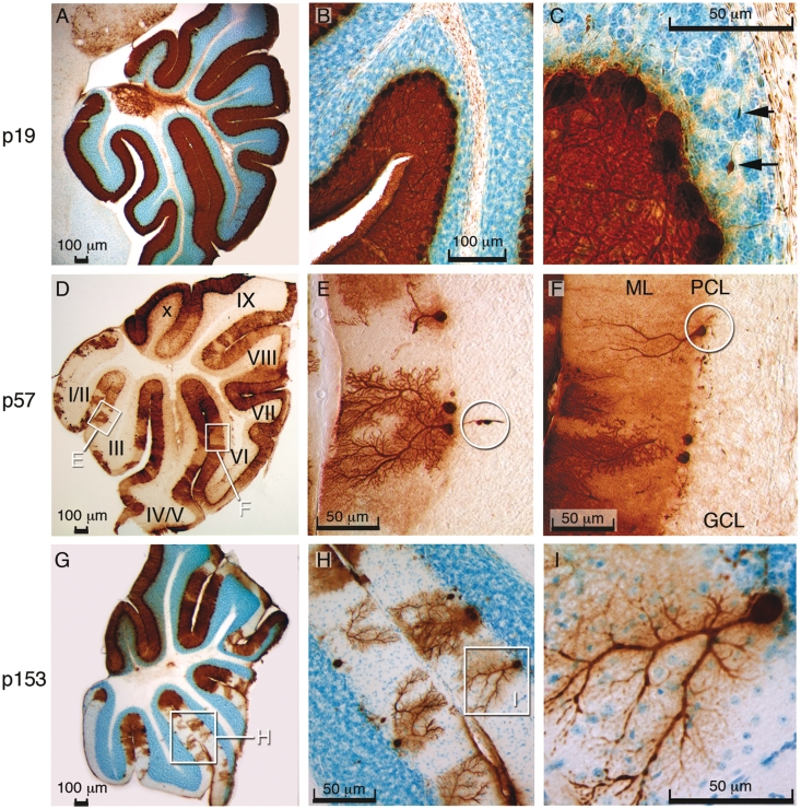
Figure 6.
Extensive and progressive loss of Purkinje cells in the mouse cerebellum. (A–C) Immunoperoxidase staining for calbindin (brown) with Nissl counterstain (blue) revealed normal cerebellar Purkinje cells in a 19-day-old (p19) Slc9a6−/Y animal. The arrows in C indicate axonal spheroids. (D) Purkinje cell degeneration, particularly noticeable in lobules I/II and III at p57. (E) Purkinje cell loss and axonal spheroids (encircled) at p57. (F) Purkinje cell that has lost all dendritic branches (ML = molecular layer; PCL = Purkinje cell layer; GCL = granular cell layer). (G) Purkinje cell degeneration is extensive with relative sparing of lobules IX and X at p153. (H) Purkinje cells in the process of losing dendritic branches. (I) Magnified view of insert in G.

