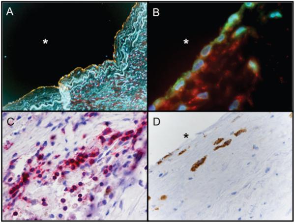
Figure 5.
Immunohistochemical staining of early, clinically inapparent human atherosclerotic lesion. A, Under stress conditions, ECs (green: von Willebrand factor) express hHSP60 (red). Yellow represents colocalization between vWF and HSP60. Blue indicates DNA. B, Adhesion molecules (green: vascular cell adhesion molecule-1) and hHSP60 (red) simultaneously expressed on the EC surface. Blue indicates DNA. The intima of the early lesion contain a higher number of T cells (C) (red: CD3) compared with macrophages (D) (brown: CD68). Original magnification: ×250 (A), ×600 (B), and ×400 (C and D). *Lumen.

