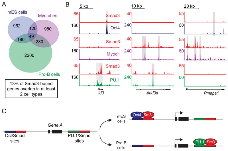
Figure 5. Smad3 can bind the same gene at different sites in different cell types.
(A) Smad3 binds a small number of genes in common between different cell types. The Venn diagram shows the overlap of genes bound by Smad3 in mES cells, myotubes and pro-B cells (Table S3). The numbers represent the total number of bound genes in each shaded area.
(B) Smad3 co-occupies the same gene but at cell-type-specific sites. Gene tracks show binding of Smad3 and Oct4 in mES cells (top), Smad3 and Myod1 in myotubes (center) and Smad3 and PU.1 in pro-B cells (bottom) for Id3, Arid3a and Pmepa1. Gray boxes highlight sites co-occupied by Smad3 and master transcription factors in each cell type. The floor is set at 3 counts.
(C) Smad3 co-occupies a fraction of genes with different master transcription factors by binding at different sites. At hypothetical Gene A one SBE (red box) is adjacent to an Oct4 site and another is adjacent to a PU.1 site. In mES cells Smad3 (Sm3) binds with Oct4 while in pro-B cells Smad3 binds with PU.1. See also Table S3.

