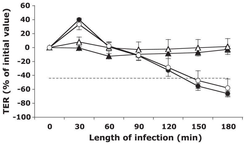
Fig. 1.
Transepithelial electrical resistance (TER) of human colon cancer-derived cell line T-84 monolayers after apical exposure to Shigella flexneri in the absence and presence of Saccharomyces boulardii over 3 h. The TER values are displayed as percentages of initial values. The data are expressed as means ± SD of triplicate samples for all conditions tested and represent 1 of at least 3 independent experiments performed with similar results. Buffer control (▲), S. boulardii only (△), S. flexneri only (●), and S. flexneri and S. boulardii together (○) are shown. The dashed line represents the point at which the barrier would likely be permeable to large molecules. No significant differences were observed in loss of TER between monolayers infected with S. flexneri only and those infected with S. flexneri in the presence of S. boulardii.

