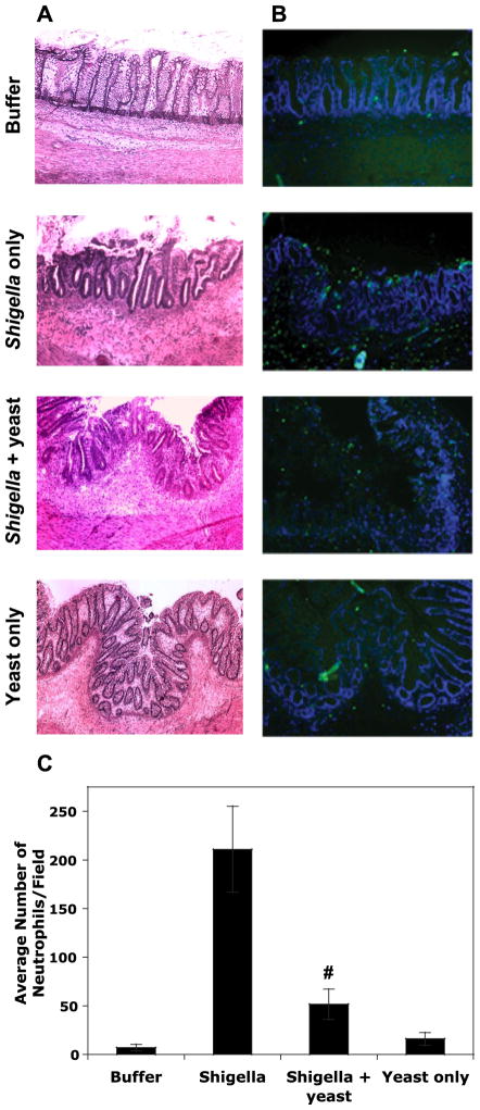
Fig. 8.
Histopathology following 24 h of infection of human fetal colonic xenografts with S. flexneri in the absence and presence of S. boulardii, sterile HBSS+, or yeast alone. A: hematoxylin and eosin-stained (see MATERIALS AND METHODS) xenotransplanted colon sections shown at ×100 magnification. B: fluorescent-stained xenotransplanted colon sections at ×100 magnification. Sections were stained with 4′,6′-diamidino-2-phenylindol, and PMNs were stained using a FITC-labeled antibody specific for PMN surface markers (Ly-6G and Ly-6C). Photographs are representative tissues for each condition (n = 3 mice for each condition). C: the average number (means ± SD) of PMNs counted in 15 fields for each of the conditions. #P < 0.01 with respect to S. flexneri with and without S. boulardii.

