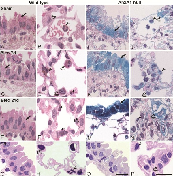
Figure 1.

Analysis of endogenous AnxA1 protein and Anxa1 gene expression during lung fibrosis. (A-H;O-P) Wild type and (I-N) AnxA1 null mice received bleomycin i.t. at time 0. The AnxA1 protein content was analyzed by immunohistochemistry. At 0 time-point, wild type mice exhibit a basal immunostain for AnxA1 protein (A and B). After 7 days post-bleomycin administration, this protein expression was greatly increased (C and D). And, after 21 days, the AnxA1 expression was reduced (E and F) in epithelial cells (arrow) and polymorphonuclear (PMN) (arrowheads) and MPC (curve arrow)(G and H). Control of immunogold reaction (CR) showing no cellular immunostaining. Anxa1 gene promoter activity was visualized by X-Gal staining reaction. AnxA1 null mice lung showing Anxa1 gene expression (I and J) on the epithelial cells (arrow), intravascular PMN (arrowhead) and MPC (curve arrow); an intense positive reaction was attained (K-N) on day 7 and 21 post-bleomycin administration. Control for LacZ reaction (CR) showing wild type mice negative to the X-Gal staining (O and P). Haematoxylin counterstain. Bars, 10 μm.
