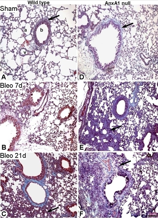
Figure 3.

Lung histopathology. (A-C) Wild type and (D-F) AnxA1 null mice lung were analyzed at 0, 7 and 21 days post-bleomycin i.t. administration as described in Material and Methods section. (A and D) Histological analysis of wild type and AnxA1 null mouse lungs showing presence of collagen in the connective tissue in the lung parenchyma (a), near the vessel (v) and bronchiole (b). (B) No major changes in the connective tissue of wild type lung parenchyma, as observed at day 7 post-bleomycin administration. (C) At day 21 post-bleomycin, the alveolar septa thicken because of a significant increase in connective tissue deposit (arrow). (E and F) In the AnxA1 null mice, the fibrosis was evident already at day 7; by day 21 there is evidence of increased connective tissue (arrows). Masson trichrome stain. Bars, 100 μm.
