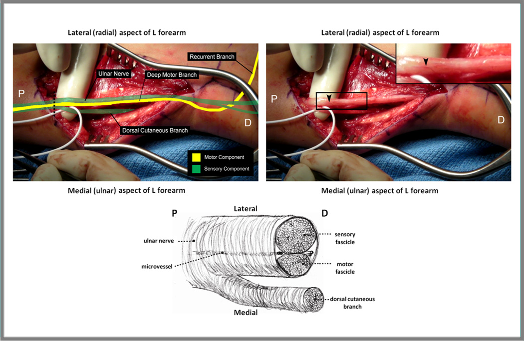
Figure 3. Ulnar nerve topography within the distal forearm.
Left: The motor (yellow) and sensory (green) fascicles should be identified at the time of transplantation to facilitate appropriate alignment during repair. Proximally, the ulnar nerve topography is sensory-motor-sensory, until the dorsal cutaneous branch separates from the remainder of the nerve approximately 10 cm proximal to the wrist crease. In order to confirm the identity of the motor fascicle, the deep motor branch should be identified within Guyon’s canal distally (not shown), then followed visually to where it joins the main sensory fascicular group proximally. Right: A prominent longitudinally oriented vessel (arrow) is frequently present, marking the natural cleavage plane between the sensory and motor fascicular groups of the ulnar nerve. Bottom: Ulnar nerve in cross section. Below the take-off of the dorsal cutaneous branch, the sensory fascicle comprises approximately 60% of the ulnar nerve, and the motor fascicle 40%. P = proximal; D = distal.Dashed line = cross section. (Adapted from Brown JM, Yee A, Mackinnon SE. Distal median to ulnar nerve transfers to restore ulnar motor and sensory function within the hand. Neurosurgery. 2009;65:966-78; with permission.)

