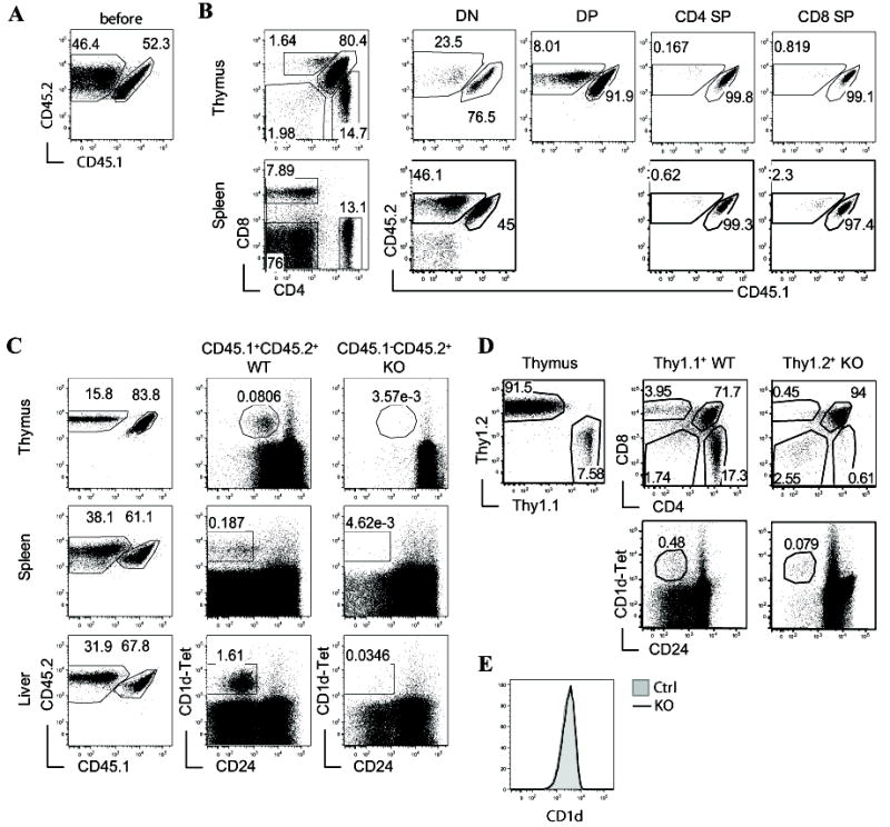
Figure 2. iNKT developmental defects in RasGRP1-/- mice are cell-intrinsic.

(A-C) Generation and analysis of sublethally irradiated TCRβ-/-δ-/- recipient mice reconstituted WT (CD45.1+CD45.2+) and RasGRP1-/- (CD45.1-CD45.2+) bone marrow (BM) at 1:1 ratio. Chimeric mice were analyzed 7-8 weeks after reconstitution. (A) Expression of CD45.1 and CD45.2 on mixed WT and RasGRP1-/- BM cells before adoptive transfer. (B) Analysis of cαβT cells and non-T cells in recipient mice. Left panels show CD4 and CD8 staining of thymocytes and splenocytes from recipient mice. Middle and right panels show CD45.1 and CD45.2 staining in the DN, DP, and SP populations based on CD4 and CD8 expression. (C) Analysis of iNKT cells in recipient mice. Left panels show CD45.1 and CD45.2 staining in the indicated organs from recipient mice. Middle and right panels show expression of CD1d-Tet and CD24 on gated CD45.1+CD45.2+ WT and CD45.1-CD45.2+ RasGRP1-/- cells. (D) Analysis of lethally irradiated WT C57B6 recipient mice reconstituted with WT (Thy1.1) and RasGRP1-/- (Thy1.2) BM cells at 1:10 ratio. Left panel, Thy1.1 and Thy1.2 staining of recipient thymocytes; Middle and right panels, CD24 and CD1d-Tet staining as well as CD4 and CD8 staining gated on Thy1.1+ and Thy1.2+ thymocytes. (E) CD1d expression on RasGRP1-/- and control CD4+CD8+ DP thymocytes. Data are representative of three (A-C) or two (D, E) experiments.
