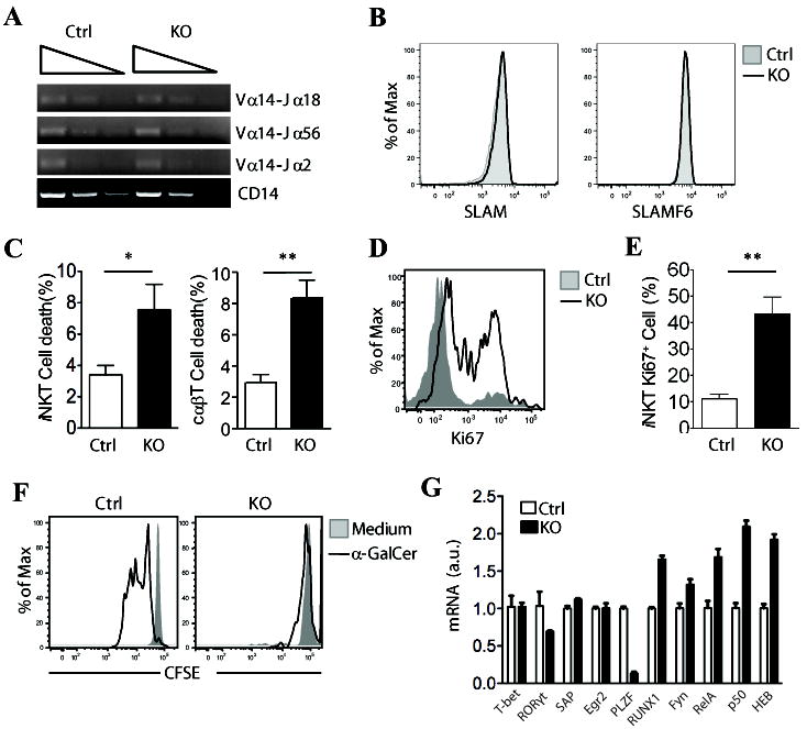
Figure 3. Increased death of iNKT cells in the absence of RasGRP1.

(A) Semi-quantitative PCR analysis of sorted CD4+CD8+ thymocytes from RasGRP1+/- and RasGRP1-/- mice with primers for Vα14-Jα2, Vα14-Jα18, Vα14-Jα56, and CD14 (loading control). (B) Expression of CD1d, SLAM (CD150), and SLAMF6 (Ly108) on CD4+CD8+ thymocytes from RasGRP1+/- and RasGRP1-/- mice. Data shown are representative of three mice per group. (C) Percentages of cell death of CD1d-Tet+TCRβ+ iNKT cells and CD1d-Tet-TCRβ+ cαβT cells from thymus (mean ± SEM, n=4). (D, E) Increased Ki67 expression in RasGRP1-/- iNKT cells. Ki67 expression in iNKT cells gated from WT and RasGRP1-/- thymocytes were determined by intracellular staining. (D) Overlay of histogram for Ki67 expression of gated iNKT cells; (E) Mean ± SEM of Ki67+ iNKT cells from WT and RasGRP1-/- thymus (n=5). (F) Impaired proliferation of RasGRP1-/- iNKT cells in response to α-Galcer stimulation in vitro. CFSE-labeled WT and RasGRP1-/- thymocytes were left unstimulated or stimulated with 125 ng/ml α-Galcer at 37°C for 72 hours. Cells were then stained for APC-CD1d-Tet and TCRβ. Overlaid histograms show CFSE levels in gated WT and RasGRP1-/- iNKT cells. (G) Real-time PCR analysis of mRNA expression of various proteins in sorted CD4+CD8+ thymocytes from RasGRP1+/- and RasGRP1-/- mice. *p<0.05; **, p<0.01 (Student t-test).
