
Figure 4.
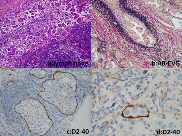
Photomicrographs. (a) A resected regional lymph node had metastatic foci composed of tumor cells with a micropapillary pattern in the case 1. (HE stain, ×200) (b) Vessel invasion by tumor cells in the case 1. (AB-EVG stain, ×100) (c, d) Tumor cells in lymphatic ducts, which were covered by D2-40 positive endothelial cells. (c, ×200, case 1; d, ×400, case 2)