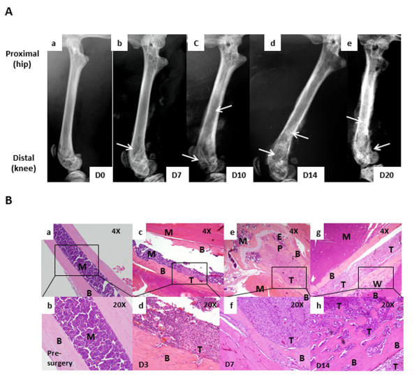
Figure 2.
Breast cancer cells when injected in the intramedullary space of the femur result into disease progression and tumor-induced bone remodeling (A). Radiographic images show normal bone with no bone destruction (a), D7 post tumor implantation with little bone loss (osteolytic remodeling; indicated by yellow arrows) observed in the shaft (b), D10 post tumor implantation with increased bone loss in the shaft and knee area (c), D13 post tumor implantation with further increased bone loss in the shaft and the knee region, cortical fractures (indicated by blue arrows) and abnormal bone growth in the shaft (osteoblastic remodeling, indicated by white arrows) (d), Histological analyses of bones (B) show control bone with intact cortical bone and marrow components (a, b), presence of tumor in the intramedullary space D3 post implantation (c, d), tumor invading into cortical bone and growing towards the epiphyseal plate of the knee by D7 post implantation (e,f), tumor infiltrating the entire intramedullary space of the bone with woven bone formation along the cortex by D13(g,h); B, cortical bone; M, bone marrow; T, tumor; EP, epiphyseal plate; WB, woven bone.

