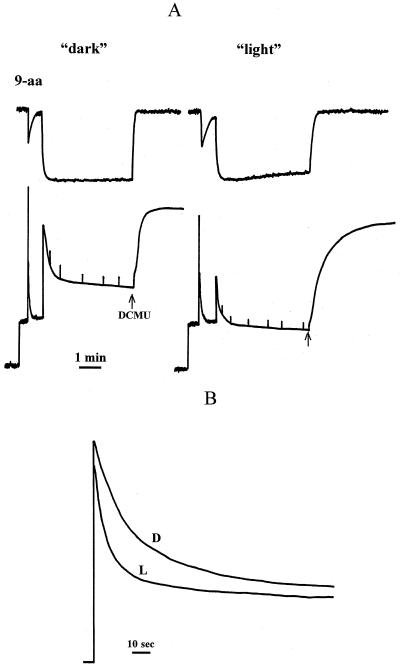
Figure 2.
A, Simultaneous measurements of chlorophyll fluorescence quenching and 9-aminoacridine (9-aa) fluorescence quenching for intact spinach chloroplasts isolated from dark-adapted (“dark”) and light-treated (“light”) leaves. Fluorescence quenching was induced by actinic light of 800 μm PAR m−2 s−1. Quenching was reversed by the addition of 5 μm of DCMU (arrow). B, Kinetics of the chlorophyll fluorescence quenching in LHCIIb diluted in buffer at pH 5.5. D and L, Samples from dark-adapted and light-treated leaves, respectively. The chlorophyll concentration of the samples was 3 μm, and the detergent concentration was 6 μm.

