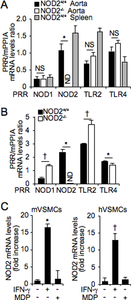
Figure 2. NOD2 mRNA is expressed in VSMCs.

Quantitative real-time PCR was performed to assess mRNA levels of PRRs in NOD2+/+ and NOD2−/− aortas (A) and VSMCs (B). Expression levels of PRRs mRNA are divided by expression of the control gene, mPPIA, and shown as a ratio of PRR/mPPIA. *p<0.05, down-regulation of gene expression in NOD2−/− vs NOD2+/+ aorta and VSMCs. †p<0.05, up-regulation of gene expression in NOD2−/− vs NOD2+/+ aorta and VSMCs. NOD2+/+ spleen was used as a positive control in (A). NS, not significant; ND, not detectable. C, Expression of NOD2 mRNA in VSMCs, after exposure to IFN-γ (10 ng/mL) or MDP (1 µg/mL), was performed by quantitative real-time PCR in mouse primary (m) and human primary (h) VSMCs. For both mouse and human NOD2 mRNA, expression was normalized by an internal control gene, either mPPIA (for mouse NOD2) or hGAPDH (for human NOD2), and shown as a fold increase. *p<0.05 vs no IFN-γ in mVSMCs. †p<0.05 vs no IFN-γ in hVSMCs. For all of the real-time PCR experiments, values are presented as mean ± SD, n=3.
