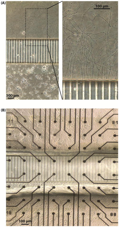
Figure 2.
(A) Unidirectional axons growing out of the microtunnels on a coverslip on DIV 12. Neurons were plated only in Well A and axons of the neurons grew through the microtunnels to reach Well B. Many axons were longer than 1 mm at this time point. The bundles of axons filled the openings of the microtunnels on the side of Well B, as shown in the enlarged view of the right panel. (B) Two neuronal networks connected with unidirectional axonal connections on an MEA. The network in Well B is connected with the network in Well A by axons throughout the microtunnels on the MEA. Cells were plated in Well A first and then in Well B ten days later. The cell bodies in both wells were separated by the microtunnels and connected by the unidirectional axons like those shown in A.

