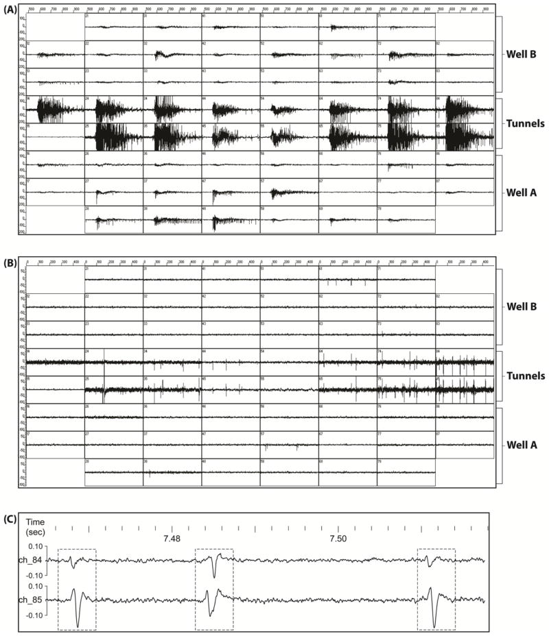
Figure 3.
Electrophysiological signals recorded from an MEA. The signals comprise two types of activities, individual spikes and synchronous bursts. (A) Spontaneous and synchronous bursts spreading throughout the whole network. (B) Individual spikes recorded during inter-burst intervals by electrodes in the region of microtunnels. Note the near simultaneity of spikes on each electrode-pair. (C) Enlarged view of the signals from an electrode-pair on channels 84 and 85. Three spike-pairs are indicated by the frames. The first and third spike-pairs are from the same unit-pair, and presumably the same axon. Each electrode signal box is +/− 200 μV and 500 msec, in A, +/− 100 μV and 500 msec in B, while the scales in C are +/−100 μV, with time in seconds.

