
Figure 1.
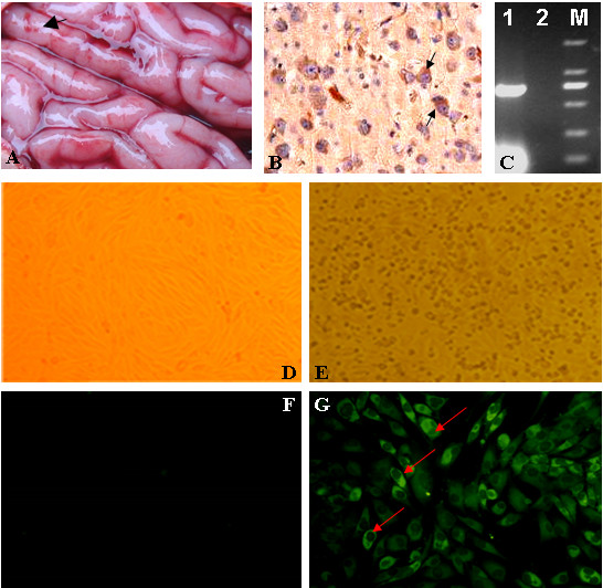
Isolation and identification of the SX09S-01 strain. (A) Lesions in the brain of piglet. Haemorrhage spots are indicated by filled arrow. (B) Immunohistochemistry (IHC) of the piglet's brain used the special monoclonal antibodies against JEV E protein. Brown cells, positive cells infected by JEV, are shown by the arrow. (C) RT-PCR results. Lane 1: 674-nucleotide fragment was amplified by special primers of the C/prM; lane 2: Negative control; lane M: DNA marker DL 2000, size of bands are 2000 bp, 1000 bp, 750 bp, 500 bp, 250 bp and 100 bp from top to bottom. (D) & (E) Cytopathic effect (CPE) of JEV in BHK-21 cells. Fig D is normal BHK-21 cells control. (F) & (G) Indirect immunofluorescent assay (IFA) with the specific monoclonal antibody against JEV E protein. Bright fluorescence cells were indicated by arrows. Figure G is normal cells control.