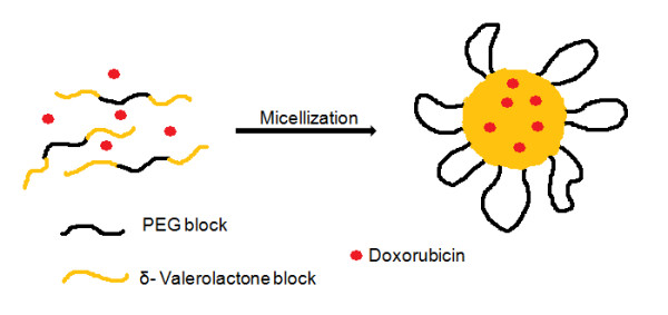
Figure 14.

Schematic illustration to represent the structure of VEVDMs. Schematic diagram showing the structure of micelles formed on DOX entrapment in VEV copolymeric micelles. The micelle is represented by a hydrophobic PVL core and hydrophilic PEG on the surface with the drug entrapped inside the hydrophobic matrix.
