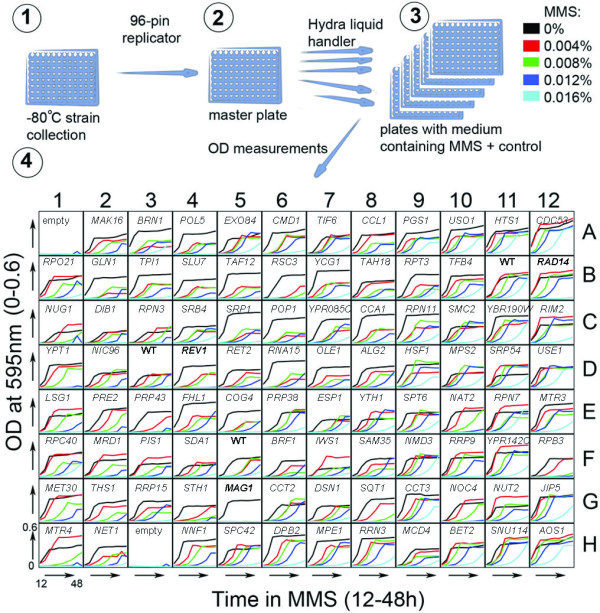
Figure 1.

Schematic of the experimental procedure. (1) Cells are kept at -80°C from where they are pin-replicated and (2) grown to stationary phase in a master plate. (3) Once in stationary phase, the cultures are robotically diluted in YPD media (using a Hydra liquid handler) containing increasing doses of MMS (0-0.016% final concentration). (4) After incubation at 25°C for 12 hours, optical densities of all cultures are measured every 4 h until 48 h post-treatment. Growth curves are plotted and data is analyzed. As an example, plate 1 from the DAmP library is shown with the name of gene deleted in each strain given above the growth curves. Every plate contains control strains (in bold), WT (B11, D3, F5), rad14Δ (B12), rev1Δ (D4) and mag1Δ (G5), as well as at least two empty wells, containing media only.
