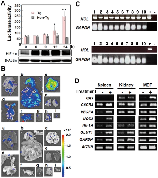
Figure 2. In vitro analysis of the HOL reporter response to HIF-1 activation.
(A) MEF cells from FVB/HOL (Tg) and FVB/N (non-Tg) mice were cultured under hypoxic conditions (1% O2) for the indicated time, and luciferase activity was measured. The experiments were performed in triplicate, and the mean luciferase activity ± SD is shown in the graph. *P<0.05, **P<0.005. (B) FVB/HOL mice were peritoneally injected with PG, and 2 h later RNA was isolated from the indicated tissues. RT-PCR analysis was performed with the isolated RNA. Glyceraldehyde-3-phosphate dehydrogenase was used as an internal control. +, indicates a transgene used as a template; −, indicates no template. The experiment was performed in triplicates, and the representative data are shown. (C) FVB/HOL (Tg) and FVB/N (non-Tg) mice were peritoneally injected with PG, and 2 h later the mice were injected with luciferin through the tail vain; 2 min later, the tissues were isolated and observed by ex vivo bioluminescent imaging. a, liver; b, intestine; c, lung; d, uterus; e, kidney; f, spleen and pancreas; g, heart; h, thymus; i, bladder.

