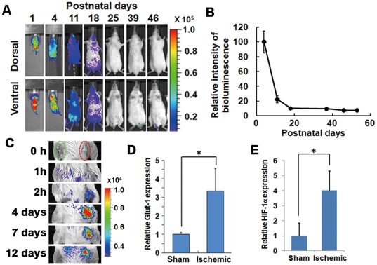
Figure 3. In vivo analysis of the HOL reporter response to HIF-1 activation.
(A, B) The HIF-1 activity after birth. FVB/HOL mice were peritoneally injected with luciferin on the indicated day after birth, and bioluminescent images were acquired 20 min later. (A) Representative images are shown. (B) Total whole body photon counts were measured 4, 11, 18, 25, 39, 46, and 53 days after birth, and the photon counts (photon counts/s/mm3) in the region of interest are shown as values normalized to that of day 4 (n = 5). Results are given as the means ± SEM. (C) The femoral artery was ligated and cut without damaging the nevus femoralis. A sham operation was performed on the ipsilateral side. Changes in bioluminescence were monitored sequentially at indicated time periods after the operation. (D, E) Seven days after the ligation surgery of the femoral vessels, RNA was isolated from the skeletal muscle of the mice legs, and semi-quantitative real-time RT-PCR was performed. GLUT1 (D) and HIF-1α (E), known as HIF-1 target genes, from the mRNA of the ischemic hindlimb muscle of the mice was quantified and normalized relative to the 18S rRNA level (n = 3). Results are given as the means ± SD. *P<0.05.

