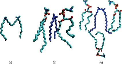
Figure 9. Snapshots selected from the MD simulations, illustrating (a) a typical conformation of the 12-10-12 molecule (the long hydrophobic spacer bending towards the interior of the bilayer), and the positioning of the (b) 12-2-12 and (c) 18-2-18 molecules relative to the neighboring phospholipid molecules.
The larger conformational freedom found close to bilayer centre observed for the short tail surfactant contrasts with the interdigitation evidence observed for the long tail surfactant.

