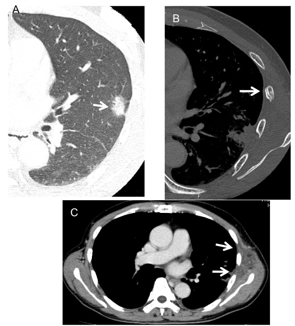
Figure 3.
A 65-year-old man with a rib fracture after SRT. A, Preradiotherapeutic thin-section CT showing a spiculated nodule with surrounding ground-glass opacity close to the chest wall (arrow). B, Twelve months later after SRT, a rib fracture is apparent (arrow). C, At 6 months after SRT, enhanced CT shows swelling of the left chest wall with an area of low attenuation (arrows).

