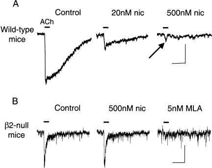
Figure 9.
Exposure to low concentrations of nicotine differentially desensitizes the fast and slow components of nicotinic currents from VTA DA neurons. (A) ACh-induced currents (1 mM ACh, 200 msec puff, horizontal line) in the absence (Control) and presence of bath-applied nicotine (20 nM or 500 nM for 20 min). Although the slow component of the current (mainly α4β2* nAChRs) was desensitized, the fast component (indicated by the notch, arrow) was not desensitized. The scale bars represent 100 pA and 0.5 sec. (B) Exposure to low concentrations of nicotine does not desensitize the fast, MLA-sensitive currents from VTA DA neurons of β2-null mice. ACh-induced currents (1 mM ACh, 200 msec puff, horizontal line) are shown from the same neuron. In the absence (Control) or presence of nicotine (500 nM for 20 min) the currents are the same. The ACh-induced currents were inhibited by 5 nM MLA, confirming that these are α7* nAChR currents. The ACh-induced currents were not inhibited by 1 μM DHβE (data not shown). The scale bars represent 50 pA and 0.5 sec. (These data are taken and adapted from Wooltorton et al. 2003.)

