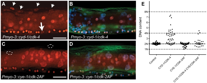
Figure 3. C. elegans Cyclin D/CDK-4 induces DNA replication in muscle.
(A-D) Detection of EdU incorporation in body-wall muscle nuclei. (A,C) EdU staining, (B,D) merged image of EdU, GFP and DAPI staining. EdU-positive nuclei are readily detectable in CYD-1/CDK-4 expressing body-wall muscle (A, arrowheads), but not in CYE-1/CDK-2AF expressing body-wall muscle (C, circles). For comparison, the arrow in (A) indicates a Pn.p cell, which completed one round of DNA replication in the presence of EdU. (E) Quantitative determination of DNA content reveals DNA replication in CYD-1/CDK-4 expressing muscle cells. Each dot indicates the DNA content, based on propidium iodide staining, of a single body-wall muscle nucleus. Pn.p nuclei in the ventral cord were used as 2n controls. L3/L4 stage larvae were stained in A-D, GFP staining reveals muscle expressed GFP::H2B and CDK-2/4::Venus.

