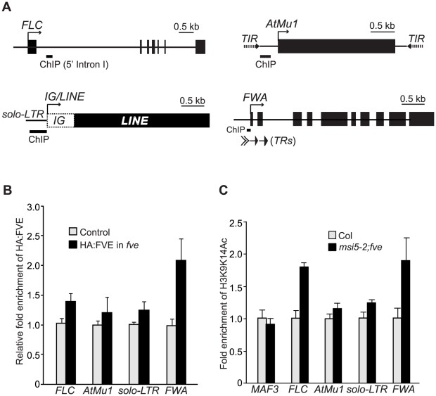
Figure 9. FVE Enriches at the Chromatin of Target Loci and Is Required for Histone Deacetylation.
(A) Schematic structures of genomic FLC, FWA, AtMu1 and IG/LINE. Solid arrows indicate the transcriptional start sites. Solid bars indicate the regions examined by ChIP. (B) HA-FVE enrichment at FLC, FWA, AtMu1 and solo-LTR. Immunoprecipitated genomic DNA was measured by real-time quantitative PCR, and subsequently normalized to the constitutively expressed TUB2 or ACTIN2 (ACT2). The fold enrichments of HA-FVE protein in the HA-FVE line over the control line (Col) are shown. Error bars indicate SD of four biological repeats. (C) Analysis of acetylated H3 (at K9 and K14) in Col and msi5-2;fve seedlings. Immunoprecipitated genomic DNA was quantified and normalized to TUB2 or ACT2. The fold enrichments of acetylated H3 in msi5-2;fve relative to Col are shown. Error bars indicate SD of two biological repeats (each quantified in triplicate). Note that MAF3, an FLC homolog, was included as a negative control to show that H3 is not hyperacetylated in a non-target gene upon loss of FVE and MSI5 function.

