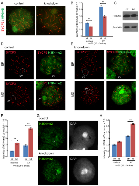
Fig. 4.
Reduced HR6A/B and increased dimethylation of histone H3 at Lys4 in Rad18 KD spermatocytes and spermatids. (A) Double immunostaining of control and Rad18 KD pachytene spermatocyte nuclei with anti-SYCP3 (red) and anti-HR6A/B (green). (B) The intensities of HR6A/B in the nucleus and on the XY body were measured with ImageJ. The intensity of HR6A/B in control nuclei was set at 1.0. Error bars indicate s.e.m. (C) HR6A/B expression in total cell extracts of testis from 4-week-old control (ctr) and Rad18 KD mice was detected on immunoblots. β-tubulin was used as loading control. (D,E) Double immunostaining of spermatocyte nuclei with anti-SYCP3 (red) and anti-H3 dimethylated at Lys4 (H3K4me2) (green) in control (D) and Rad18 KD mice (E). EP, early pachytene; MD, mid diplotene. The XY body is shown in the white circle. (F) The intensities of H3K4me2 in the nucleus and on the XY body from 4-week-old and 19-week-old mice were measured using ImageJ. The intensity of H3K4me2 in control nuclei was set at 1.0. Error bars indicate s.e.m. (G) Immunostaining of spermatid nuclei with anti-H3K4me2 (green) in Rad18 KD and control mice. Arrows indicate sites of the accumulation of H3K4me2. (H) The intensities of H3K4me2 in spermatid nuclei and on the X or Y chromosomes were measured as described in F. (B,F,H) Blue and red bars indicate control (ctr) and Rad18 KD, respectively. **P<0.01 (Mann–Whitney U-test).

