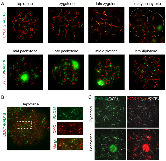
Fig. 7.
Accumulation of RAD18 at IR-induced DSBs. (A) Localization of RAD18 in spermatocyte nuclei of wild-type mice irradiated with IR at 4Gy. Two hours later, spread spermatocyte nuclei were prepared and double-stained with anti-SYCP3 (red) and anti-RAD18 (green) antibodies. The white line in the early pachytene image indicates the boundary of the nucleus. (B) Double immunostaining of irradiated (as in A) leptotene nucleus with anti-DMC1 (red) and anti-RAD18 (green). On the right, an enlarged region is shown with separate red and green images, to better visualize foci that do and do not colocalize. (C) Triple immunostaining of irradiated (as in A) zygotene and pachytene nuclei with anti-H2AK119ub1 (red), anti-RAD18 (green) and anti-SYCP3 (white).

