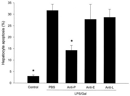
FIG. 7.
Apoptosis of hepatocytes 6 h after treatment with a combination of LPS (10 μg) and Gal (18 mg) and monoclonal antibodies against P-selectin (RB40.34, 40 μg; anti-P), E-selectin (10E9.6, 40 μg; anti-E), or L-selectin (Mel-14, 40 μg; anti-L) or PBS. Control animals received PBS. Hepatocyte apoptosis is given as the percentage of observed hepatocyte nuclei with morphological signs of apoptosis, i.e., chromatin condensation and fragmentation, after administration of the fluorochrome Hoechst 33342. Data represent means ± SEM. Asterisks indicate significant differences versus challenge with LPS-Gal plus PBS (P < 0.05).

