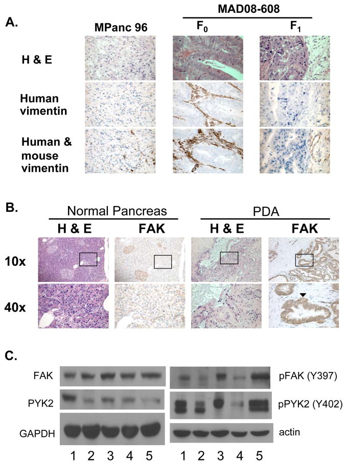
Figure 1.
FAK expression in PDA. A, Representative photomicrographs are shown from tumor sections of mice bearing MPanc-96 or MAD08-608 F1 tumors and from the original patient tumor, designated MAD08-608 F0. Human-specific vimentin antibody (middle panel) demonstrates human-derived stromal tissue. Staining of tumors with an antibody recognizing both mouse and human-derived vimentin (bottom panel) demonstrated staining of stromal tissue in each of the tumor samples. B, The staining for FAK observed throughout the PDA sections was most intense in the malignant ductal cells (arrowhead). C, The level of FAK and PYK2 was determined by Western blotting of human PDA cell lines (1, BxPC-3; 2, L3.6pl; and 3, MPanc-96) the patient-derived tumor, MAD08-608 (4) and HPSCs (5). Parallel analysis was carried out to detect phosphotyrosine containing FAK and PYK2.

