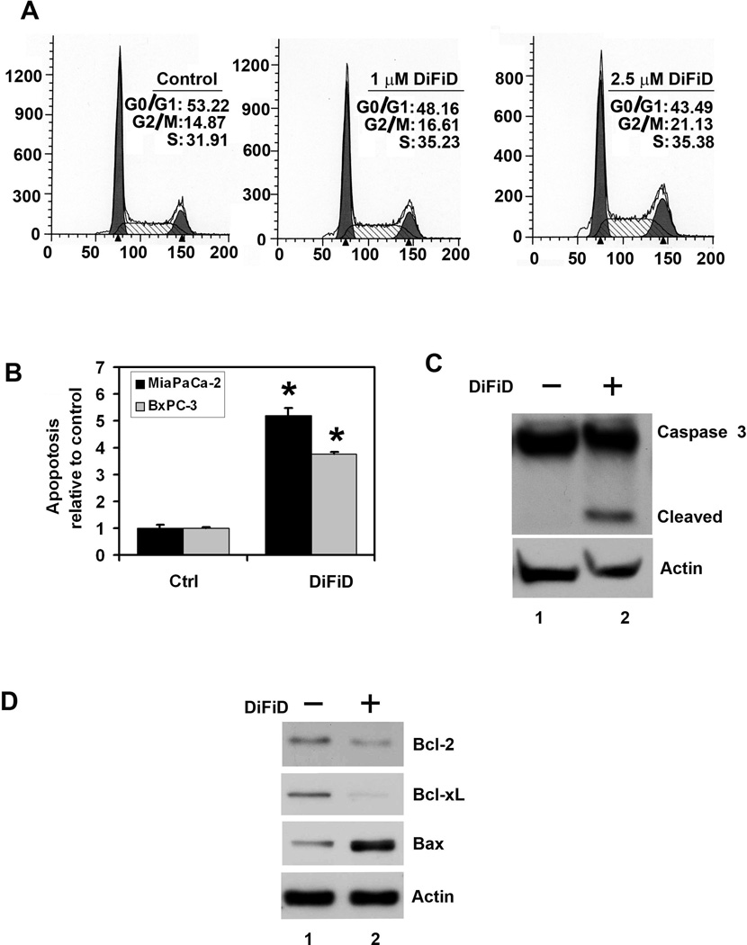
Figure 2. DiFiD induces cancer cell apoptosis.
A, Cell cycle analysis of DiFiD treated cells. MiaPaCa-2 cells were treated with up to 2.5 µM DiFiD for 24 h and examined by flow cytometry following propidium iodide staining for DNA content. DiFiD treatment leads to increased number of cells in the G2/M phase. Graphs are representative of data collected from three experiments. B, DiFiD treatment induces apoptosis in MiaPaCa-2 and BxPC-3 cells. The cells incubated with 1 µM DiFiD for 24 h and analyzed for apoptosis by caspase 3/7 activation. DiFiD treatment increased the number of apoptotic cells compared to untreated controls (*P <0.05). Results are from three independent experiments. C, DiFiD induces caspase 3, an apoptosis mediator. Lysates from MiaPaCa-2 cells incubated with 1 µM DiFiD were analyzed by western blotting for caspase 3 protein levels using rabbit anti-caspase 3 antibody. DiFiD treated cells shows cleaved (activated) caspase 3 while untreated cells have no cleaved caspase-3. D, DiFiD reduces expression of anti-apoptotic proteins Bcl-2 and Bcl-xL in treated cells when compared to untreated cells. Lysates from MiaPaCa-2 cells incubated with 1 µM DiFiD were analyzed by western blotting for Bcl-2, Bcl-xL, and Bax proteins. Both Bcl-2 and Bcl-xL were reduced while Bax expression was increased following DiFiD treatment.

