
Fig. 5.
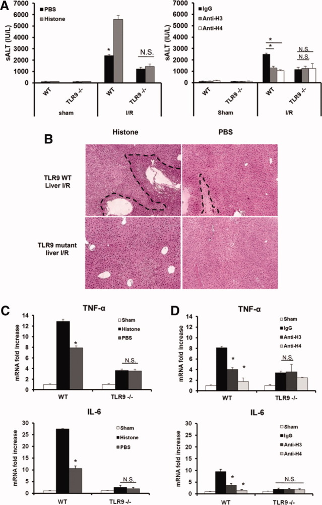
Extracellular histones mediate hepatic I/R injury through TLR9. (A) Serum ALT levels in TLR9 mutant and WT mice after I/R with either anti-histone antibodies or exogenous histone administration. Data represent the mean ± SE (n = 4-6 mice per group). *P < 0.05. (B) Hematoxylin and eosin–stained liver sections (original magnification ×100). Images are representative liver sections from six mice per group. The dashed line indicates the necrotic area. (C) Hepatic TNF-α and IL-6 mRNA in TLR9 mutant and WT mice after histone administration. (D) Hepatic TNF-α and IL-6 mRNA expression after histone neutralization. Results are expressed as the relative increase of mRNA expression compared with sham-treated animals. Data represent the mean ± SE (n = 4-6 mice per group). *P < 0.05. N.S., not significant.