
Fig. 1.
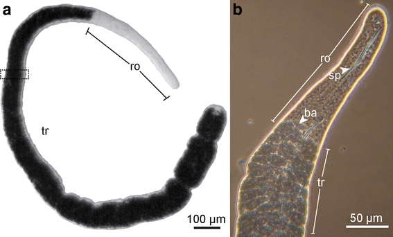
Habitus of Paracatenula cf. polyhymnia. a Light microscope micrograph, dotted rectangle represents area of serial section. b Phase contrast micrograph of the anterior region. tr trophosome region, ro rostrum, sp spicule, ba bacteria
Official websites use .gov
A
.gov website belongs to an official
government organization in the United States.
Secure .gov websites use HTTPS
A lock (
) or https:// means you've safely
connected to the .gov website. Share sensitive
information only on official, secure websites.
Habitus of Paracatenula cf. polyhymnia. a Light microscope micrograph, dotted rectangle represents area of serial section. b Phase contrast micrograph of the anterior region. tr trophosome region, ro rostrum, sp spicule, ba bacteria