
Fig. 3.
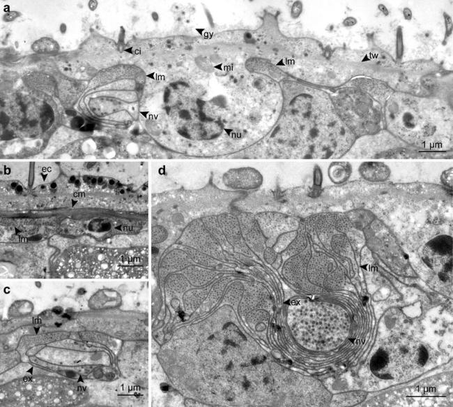
Detailed TEM micrographs of the body wall, showing epidermal cells, muscles, nerves and the dorsal cord in cross-sections. a Cross-section of a epidermal cell. b Circular muscles below the epidermis. c Nerve cell associated with longitudinal musculature. d Cross-section of the dorsal cord. cm circular muscle, ci cilium, ec epidermis cell, ex extension, gy glycocalyx, lm longitudinal muscle, mi mitochondrion, nu nucleus, nv nerve cell with vesicles, tw terminal web