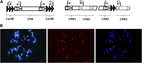
Figure 1 .
Centromeric retrotransposons (RTc) amplification and confirmation. (A, left) Single insertion of centromeric retrotransposon (CRB) is amplified from within an array of centromere satellite repeats (CentB). (Right) Nested insertion or doubled insertion of retrotransposons is amplified from within an interretrotransposon region, where the PCR products would be expected either to contain CentB in a doubled insertion interretrotransposon or not contain CentB in nested insertion. (B) BAC–FISH analysis using a Tapidor BAC clone (JBnB029F04) containing the RTc sequence used to design primers for the RTc markers. From left to right are chromosomes stained by 4′,6-diamidino-2-phenylindole in blue, FISH signals derived from BAC clone (red), and merged image. A total of 36 of 38 chromosomes with centromeres are visible in the cell. Major signals were localized in the centromere, indicating that the RTc markers were centromere specific.

