Figure 1—
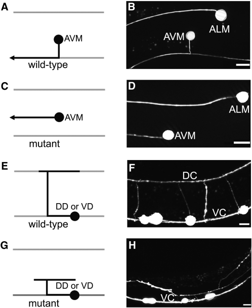
Mutations cause axon migration phenotypes. Left column, schematic diagram of the AVM neuron (A and C) and the DD and VD neurons (E and G); right column, fluorescence photomicrographs of larvae bearing a mec-4::gfp transgene that expressed GFP in the AVM neuron (B and D) or a unc-47::gfp transgene that expressed GFP in the DD and VD motor neurons (F and H). In each panel, left is anterior and up is dorsal. Bars, 10 µm. (A and B) In wild-type larvae the AVM neuron is located on the lateral right side of the C. elegans body wall. During the L2 stage, the AVM axon is guided ventrally to the ventral nerve cord where it turns and migrates anteriorly to the nerve ring. (C and D) In mutant larvae that have axon migration defects caused by loss of UNC-6 or SLT-1 guidance signaling, the AVM axon often fails to migrate ventrally and instead travels anteriorly. (E and F) In wild-type larvae the DD and VD motor neurons send processes along the ventral nerve cord and circumferentially along the body wall to the dorsal midline. DC, dorsal nerve cord; VC, ventral nerve cord. (G and H) In mutant larvae that have axon migration defects caused by loss of UNC-6 guidance signaling the DD and VD axons wander across the body wall and rarely reach the dorsal midline.

