
Figure 2.
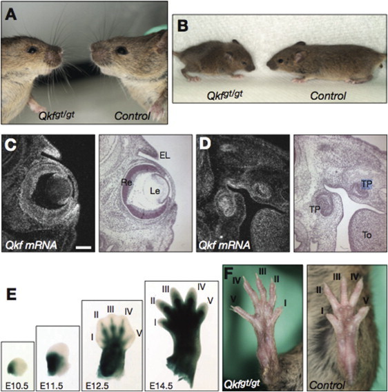
Morphology of Qkfgt/gt Mutant Mice and Expression of Kat6b mRNA in Tissues Affected in SBBYSS Individuals
(A) Facial features including small eyes and upturned nose in a 10-month-old male Qkfgt/gt mutant mouse compared to a same-sex wild-type littermate control.
(B) External appearance of a 3-week-old male Qkfgt/gt mutant mouse and a male littermate control.
(C) Qkf mRNA distribution in E15.5 eyes and eyelids. Precipitated silver grains representing Qkf mRNA appear white in the darkfield image; the brightfield image is in the adjacent panel.
(D) Qkf mRNA distribution in E15.5 tooth primordia.
(E) β-galactosidase reporter staining (blue) indicates high-expression activity of the Qkf locus in the developing hindlimb at the stages indicated.
(F) Feet of a 3-week-old male Qkfgt/gt mutant mouse and a male littermate control. Note the long and slender appearance of the Qkfgt/gt foot and digits. The following abbreviations are used: I to V, digits I to V of the hindlimb; EL, eyelids; Le, lens; To, tongue; TP, tooth primordium; Re, retina. The scale bars (C and D) equal 133 μm. Embryo recovery, histological processing and sectioning, radioactive in situ hybridization, and β-galactosidase staining were performed as previously published.10, 20, 21