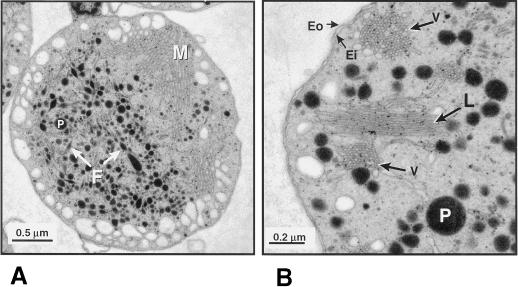
Figure 1.
Electron micrograph images of isolated red bell pepper chromoplasts. A, Representative chromoplast showing internal membranes (M), osmiophilic plastoglobules (P), and fibrils (F). B, Detail of chromoplast internal membrane compartments. Note the inner and outer envelope membranes (Ei and Eo) and the internal membranes organized into vesicles (V) and lamellae (L).

