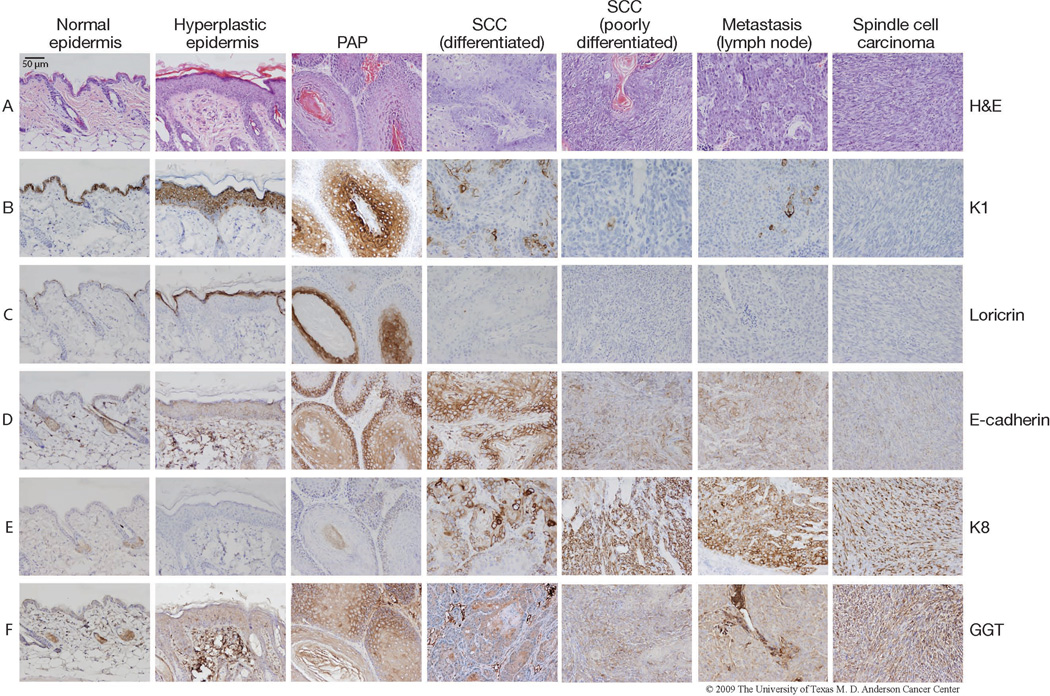
Figure 2. Expression of several marker proteins in each sequential stage of skin carcinogenesis in mice.
Tumor tissue was harvested from FVB mice that had undergone two-stage skin carcinogenesis initiated by DMBA and promoted by TPA. The tumors as well as hyperplastic dorsal skin from between tumors and untreated ventral skin were fixed in formalin and embedded in paraffin for immunohistochemical analyses. Representative images of normal epidermis, hyperplastic epidermis, papilloma (PAP), differentiated SCC, poorly differentiated SCC, lymph node metastasis and spindle cell carcinoma are shown. The lymph node metastasis was harvested from the same mouse bearing the poorly differentiated SCC. Immunostaining using the following antibodies was performed at the Histology Core at M.D. Anderson Cancer Center Science Park-Research Division as previously described24: Loricrin (Covance, Princeton, NJ), K1 (Covance), K8 (Origene, Rockville, MD), K15 (Covance), E-cadherin (Santa Cruz), and GTT (Abcam, Cambridge, MA). All mice were handled in accordance with institutional and national regulations.

