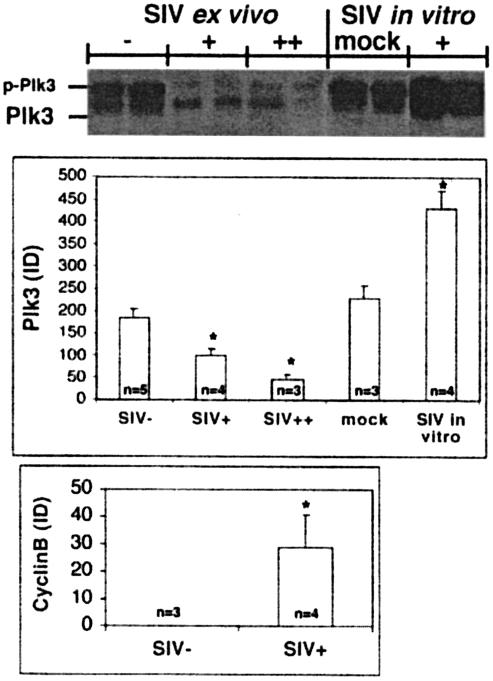
FIG. 1.
Effect of in vivo and in vitro SIV infection on expression of Plk3. Lysates from CD4+ T cells obtained from SIV-negative (SIV−) or SIV-positive (SIV+) RM with viral loads of <106 (+) or >107 (++) copies/ml and from in vitro mock-infected (mock) and SIVmac251-infected CD4+ T cells (+) were assayed for the expression of Plk3 by Western blotting. p-Plk3 denotes a slower-migrating species of Plk3, presumably the phosphorylated form. Representative data from two animals from each group (consisting of at least three animals) are shown. Results of Western blot analysis of Plk3 and cyclin B expression were quantified by densitometry, normalized to the levels of β-actin, and expressed as average integrated density (ID). Error bars represent SD of values obtained from each experimental group containing samples from n animals, and an asterisk denotes a statistically significant difference (P < 0.001) from the SIV-negative samples.

