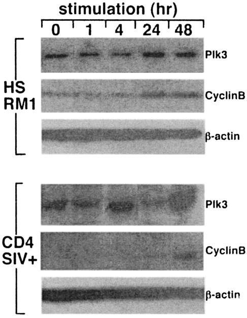
FIG. 6.
Plk3 expression in CD4+ T cells from SIV-infected RM exhibits aberrant kinetics after stimulation. HSRM1 cells and CD4+ T cells from SIV-infected RM were stimulated with anti-CD3-anti-CD28 antibody-coated beads for the indicated time intervals and assayed for the expression of Plk3 and cyclin B; β-actin expression was used as a control for the equivalent loading. For primary CD4+ T cells representative data from a group of three animals are shown.

