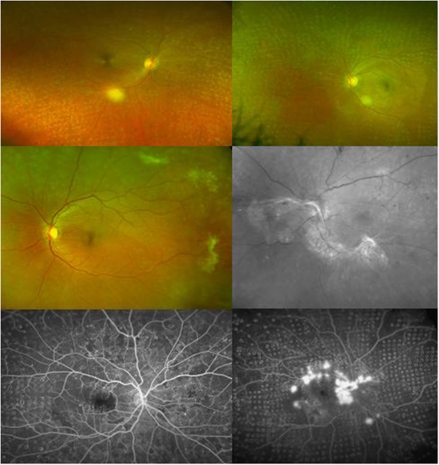
Figure 1.
Wide-field Optos imaging of PDR treated with 20 ms PRP. Upper left: Colour image showing extensive PRP coverage and complete regression of moderate PDR, and fibrosis of NVE in the peripapillary area. Upper right: Colour image of extensive PRP coverage and complete regression of mild PDR, with involution of a nasal NVE complex. Middle left: Complete regression and fibrosis of multiple NVE complexes. Middle right: Red-free image showing a PRP laser non-responder with the presence of an extensive tractional retinal detachment arising from NVD. Lower left: Optos fluorescein angiogram showing complete regression of moderate PDR and absence of NVE leakage. Lower right: Optos fluorescein angiogram showing an example of severe PDR after a single session of PRP, with active leakage from multiple NVE and NVD.

